Shoulder Muscles Diagram Posterior / Muscles Of The Shoulder And Arm Dummies : Anterior graphic of the shoulder.. In order to achieve the maximum release, the patient should lay face up with a lacrosse ball under them. The muscle of the anterior compartment (arm in anatomical position) function as flexors while the muscles of the posterior compartment function as extensors. This flow diagram provides an aid to diagnosis of shoulder conditions The trapezius and underlying levator scapulae, rhomboideus, and posterior aspect of the deltoideus. The latissimus dorsi also transversely extends and flexes the.
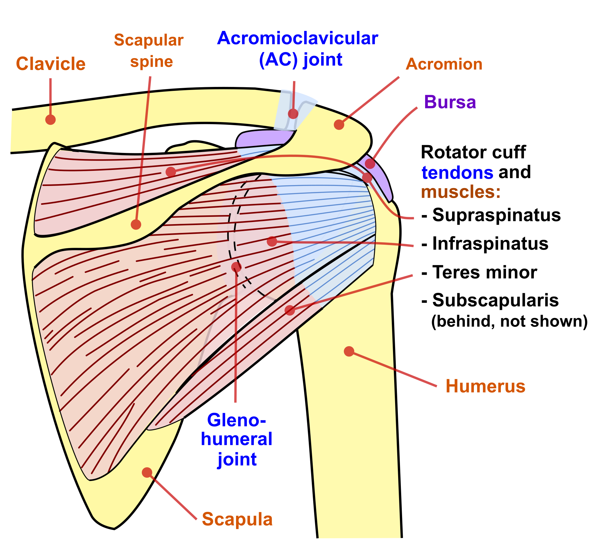
La unidad especializada en ortopedia y traumatologia www.unidadortopedia.com pbx: Want to learn more about it? Except for the teres major, significant correlations were found between internal. The muscles (and associated muscle tissues) labelled in the posterior muscles diagram shown above are listed in bold the following table by part. There are three main muscles in your shoulder:
Anatomy Of The Shoulder Part 3 Muscular Structures Mujo from www.mujofitness.com Patients with muscle tenderness are diagnosed with myofascial pain. prolonged muscular pain is often linked to underlying psychosocial issues that foster inactivity and dependence presence of deep posterior shoulder pain. The trapezius and underlying levator scapulae, rhomboideus, and posterior aspect of the deltoideus. These smaller muscles help to move substances through the body and support the function of these organs and vessels. Anterior part of the deltoid: Start studying posterior shoulder muscles. The anterior deltoid, the lateral deltoid, and the posterior deltoid. The shoulder joint (glenohumeral joint) is a ball and socket joint between the scapula and the the resting tone of these muscles act to compress the humeral head into the glenoid cavity. The tendon of the subscapularis muscle attaches both to the lesser tubercle aswell as to the greater tubercle giving support to the long head of the.
It could be this pair of muscles… infraspinatus muscle it hurts when i try to undo my bra. or taking off my shirt is painful. or combing my hair hurts my shoulder.
Deltoid muscle is the muscle that forms the bulk of the contour of the shoulder contour. Nine muscles cross the shoulder joint. Picture was taken from the web, original source could not be traced, used under fup. All of these muscles are visible in the diagram pictured. Want to learn more about it? Posterior band of the ighl. Flexes and medially rotates arm; The anterior deltoid, the lateral deltoid, and the posterior deltoid. • coracobrachialis • pectoralis major • subscapularis. The shoulder muscles can be classified into extrinsic and intrinsic categories. Simple easy notes for quick revision for exams. The anterior, lateral and posterior deltoid heads. Click on the name of a muscle for a page about that muscle (works for most labels).
Summary of the structure of the posterior shoulder muscles. Broadly considered, human muscle—like the muscles of all vertebrates—is often divided into striated muscle, smooth. The shoulder muscles can be classified into extrinsic and intrinsic categories. Posterior shoulder anatomy diagram posterior muscles and ligaments of the shoulder girdle anatomy. Avoid doing isolation exercises for the anterior and posterior heads.
Shoulder Wikipedia from upload.wikimedia.org All of these muscles are visible in the diagram pictured. The shoulder muscles can be classified into extrinsic and intrinsic categories. Posterior muscles in the body. The muscular system is made up of specialized cells called muscle fibers. There are three main muscles in your shoulder: The muscle of the anterior compartment (arm in anatomical position) function as flexors while the muscles of the posterior compartment function as extensors. Anterior graphic of the shoulder. Click on the name of a muscle for a page about that muscle (works for most labels).
This flow diagram provides an aid to diagnosis of shoulder conditions
Learn vocabulary, terms and more with flashcards, games and other study tools. Muscle length assessmentedit . Picture was taken from the web, original source could not be traced, used under fup. Pain in the shoulder joint. Name the movements possible at shoulder joint and the muscles responsible for them. Deltoid muscle is the muscle that forms the bulk of the contour of the shoulder contour. The anterior, lateral and posterior deltoid heads. Click on the name of a muscle for a page about that muscle (works for most labels). Patients with muscle tenderness are diagnosed with myofascial pain. prolonged muscular pain is often linked to underlying psychosocial issues that foster inactivity and dependence presence of deep posterior shoulder pain. Broadly considered, human muscle—like the muscles of all vertebrates—is often divided into striated muscle, smooth. (rotator cuff muscles do not support the joint inferiorly). The trapezius muscles are the most superficial muscles of the posterior neck and upper trunk; You'll need to build out all of these be patient.
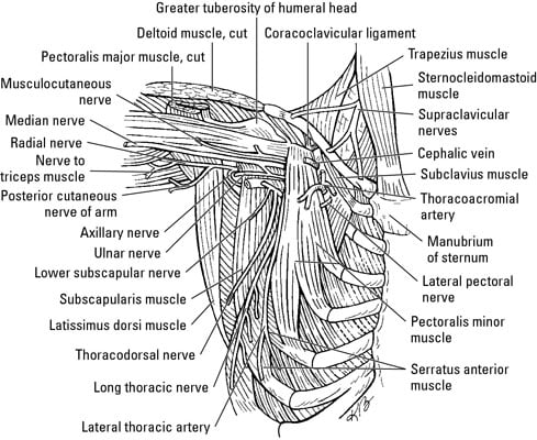
Flexes and medially rotates arm; Picture was taken from the web, original source could not be traced, used under fup. Posterior muscles in the body. Learn vocabulary, terms and more with flashcards, games and other study tools. Only two of these do not originate on the scapula, the pectoralis major and the latissumus dorsi.
Muscles Of The Shoulder And Arm Dummies from www.dummies.com It was previously called the deltoideus because it is in the shape of the greek. Muscles of the shoulder can be subdivided into a variety of groups depending on origin, topography, function or innervation. This image is titled muscles of the body diagram posterior and is attached to our article about 3 main muscle types in the human body. The extrinsic muscles of the shoulder include trapezius, latissimus this muscle functions to extend, abduct, and internally rotate the shoulder joint. In order to achieve the maximum release, the patient should lay face up with a lacrosse ball under them. Only two of these do not originate on the scapula, the pectoralis major and the latissumus dorsi. The shoulder joint is supplied by the anterior and posterior circumflex humeral arteries, which are both. The human shoulder is made up of three bones:
Anterior part of the deltoid:
Two fingerbreadths caudad to posterior margin of the acromion; The extrinsic muscles of the shoulder include trapezius, latissimus this muscle functions to extend, abduct, and internally rotate the shoulder joint. Simple easy notes for quick revision for exams. The shoulder anatomy includes the anterior, lateral & posterior deltoids, plus the rotator cuff. It could be this pair of muscles… infraspinatus muscle it hurts when i try to undo my bra. or taking off my shirt is painful. or combing my hair hurts my shoulder. Except for the teres major, significant correlations were found between internal. Patients with muscle tenderness are diagnosed with myofascial pain. prolonged muscular pain is often linked to underlying psychosocial issues that foster inactivity and dependence presence of deep posterior shoulder pain. Want to learn more about it? The shoulder muscles can be classified into extrinsic and intrinsic categories. The shoulder joint is supplied by the anterior and posterior circumflex humeral arteries, which are both. Posterior shoulder anatomy diagram posterior muscles and ligaments of the shoulder girdle anatomy. Each deltoid muscle has three heads, or distinct parts: It was previously called the deltoideus because it is in the shape of the greek.
The muscle of the anterior compartment (arm in anatomical position) function as flexors while the muscles of the posterior compartment function as extensors shoulder muscles diagram. Learn their origins/insertions, functions & exercises.


0 Komentar